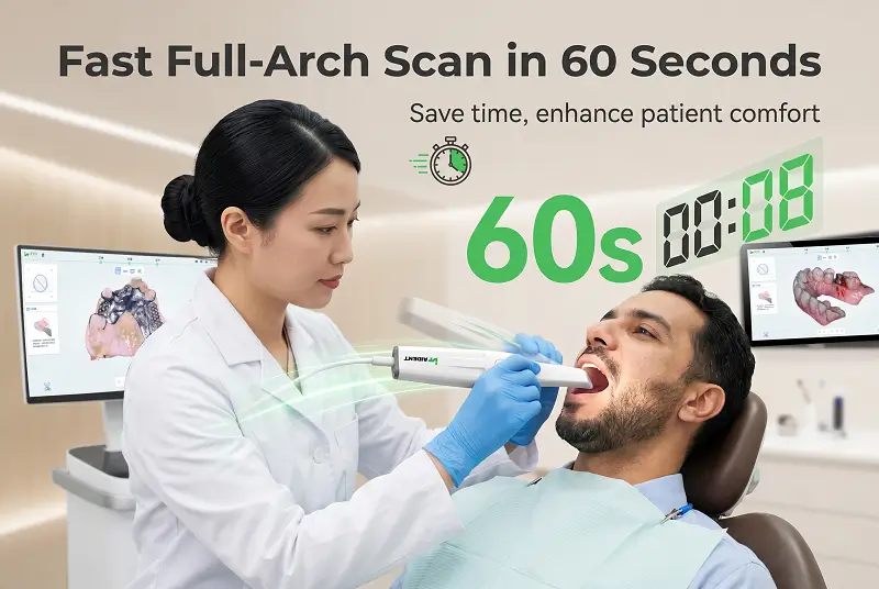
Accurate digital impressions are the foundation of every successful Scan → Design → Print workflow. Yet certain clinical conditions - dark tooth surfaces, moisture, saliva, blood, or highly reflective restorations - still challenge many intraoral scanners. Understanding how to manage these situations significantly improves scan quality, reduces chair time and minimizes the need for rescans.
This guide provides practical, field-tested techniques for scanning dark teeth and wet environments, along with the scanner features that make these cases more predictable.
Why Dark Teeth and Moisture Cause Scanning Problems
Most intraoral scanners rely on optical principles (structured light, confocal, or similar). Challenges arise when:
Dark or highly absorbent surfaces (deep stains, dark composites, metal, or tetracycline-stained teeth) reduce reflected light, leading to missing data or noise.
Moisture, saliva or blood creates reflective or refractive surfaces that confuse the optical system and produce artifacts or holes in the mesh.
Fogging on the scanner tip during intraoral use further degrades image quality, especially in humid conditions or with prolonged scanning.
Traditional workarounds such as applying powder can help in some cases, but they add time, create mess, and reduce patient comfort - especially for children or gag-sensitive patients.
Proven Clinical Techniques
1. Control Moisture First
Use high-volume suction and cotton rolls or absorbent pads strategically.
Isolate the quadrant or arch as much as practical before starting the scan.
For very wet fields, quickly dry the surfaces with air, then begin scanning immediately while the field remains relatively dry.
Work in smaller sections rather than attempting a continuous full-arch scan in a persistently wet environment.
2. Optimize Scanning Strategy for Dark Surfaces
Maintain the recommended distance and angulation; avoid extreme angles that reduce light return.
Move the scanner tip more slowly over dark or absorbent areas to allow the system to capture sufficient data.
Use overlapping passes and approach dark surfaces from multiple directions.
If the software shows persistent holes, re-scan the specific region after slightly changing the tip orientation or drying the surface again.
3. Manage Soft Tissue and Bleeding
Control hemorrhage before scanning whenever possible.
Retract soft tissue gently but consistently so that margins remain visible.
For subgingival margins, ensure the area is as dry and accessible as the clinical situation allows.
4. Leverage Scanner Features That Help in Difficult Conditions
Modern powder-free scanners with advanced optical and software capabilities handle these challenges more effectively than earlier generations.
Key helpful features include:
Powder-free operation - eliminates the need for contrast powder, saving time and improving patient experience.
Intelligent anti-fog technology - heated tips or active anti-fog systems keep the optical window clear during extended intraoral use.
True-color rendering - assists the clinician in visually confirming that critical areas (margins, dark surfaces) have been adequately captured.
Large depth of field - maintains focus across a wider range of distances, useful when access is limited or tissue is uneven.
Fast acquisition speed and intelligent software filtering - reduces the impact of momentary moisture or movement.
5. Software and Post-Scan Checks
Review the live or completed scan carefully for holes, especially on dark teeth and proximal surfaces.
Use the scanner software's gap-filling or AI-assisted tools only as a supplement - never as a substitute for adequate data capture.
If critical areas remain incomplete, rescan selectively rather than restarting the entire arch.
Recommended Scanner Characteristics for Challenging Cases
When selecting or evaluating an intraoral scanner for daily use that includes difficult cases, prioritize:
| Feature | Clinical Benefit |
|---|---|
| Powder-free | Faster workflow, better patient comfort |
| Intelligent anti-fog | Clear optics in humid or prolonged scanning |
| True-color real-time view | Easier visual verification of dark/wet areas |
| Large depth of field | More forgiving of distance variation |
| Lightweight tip & balanced design | Reduced operator fatigue during careful scanning |
| Open STL/PLY/OBJ export | Easy transfer to design and printing systems |
Aident AI-30 Intraoral Scanner incorporates these practical features:
Powder-free operation
Intelligent heating anti-fog
True-color real-time rendering
Depth of field 0–25 mm
Weight 156–198 g
Full-arch accuracy ≤10 μm
Typical full-arch scan time 20–60 seconds
Fully open output formats (STL / PLY / OBJ)
These characteristics help clinicians manage dark teeth and moist environments more efficiently while maintaining the high accuracy required for subsequent design and 3D printing steps.
Workflow Integration Tips
Train assistants on moisture control and basic scanning strategy so the dentist can focus on critical margins.
Establish a consistent sequence (e.g., occlusal → buccal → lingual) that can be adapted when conditions deteriorate.
Keep spare tips and a simple drying protocol ready for challenging patients.
Archive both the original scan and any repaired or optimized files for future reference.
Conclusion
Dark teeth and wet environments no longer need to be major obstacles to high-quality digital impressions. With proper moisture control, deliberate scanning technique, and a powder-free scanner equipped with effective anti-fog and true-color capabilities, clinicians can capture accurate data even in less-than-ideal conditions.
Clean digital impressions form the starting point of reliable personalized dentistry - from temporary crowns and surgical guides to orthodontic models and custom appliances. An open, accurate scanning foundation makes the entire Scan → Design → Print chain more predictable.
Aident3D's AI-30 intraoral scanner is designed to support exactly these demanding clinical realities while remaining lightweight, fast and fully open.
Ready to improve scanning consistency in difficult cases?
Request a demonstration or quote:
→ https://www.aident3d.com/inquiry
WhatsApp: +86 19311417410
Email: dongjiahao@aident.cc
Better scans in real clinical conditions. Better outcomes downstream.