Periodontal disease management requires consistent, accurate monitoring of soft-tissue levels, inflammation, recession, and response to treatment over time. Traditional methods-periodontal charting, clinical photographs, and physical study models-have limitations in precision, storage, and patient communication. In 2026, digital intraoral scanning combined with 3D printed models offers a more reliable, efficient, and patient-friendly alternative for ongoing periodontal care.
This article explains how digital scanning supports periodontal monitoring, how 3D printed models enhance clinical and educational applications, and how clinics can implement a practical digital workflow.
Limitations of Traditional Periodontal Monitoring
Conventional approaches face several challenges:
Manual periodontal charting is time-consuming and subject to inter-examiner variability
Clinical photographs provide only two-dimensional information and can be affected by lighting and angulation
Physical study models are bulky, fragile, difficult to store long-term, and degrade over time
Comparing sequential records accurately is often imprecise
Patient understanding of disease progression or treatment results remains limited
These limitations can reduce the effectiveness of long-term monitoring and patient engagement.
Role of Digital Intraoral Scanning in Periodontal Monitoring
High-accuracy intraoral scanners capture detailed three-dimensional information of both hard and soft tissues in a single, rapid procedure:
Precise recording of gingival margins, recession levels, and soft-tissue contours
True-color imaging that helps document inflammation and tissue health
Fast, powder-free scanning that improves patient comfort-especially important for patients with sensitive tissues
Open file formats (STL / PLY / OBJ) that allow easy storage, retrieval, and comparison of sequential scans
Ability to overlay or compare scans over time to visualize changes in tissue levels or treatment response
When scan accuracy reaches ≤10 μm, small but clinically relevant changes become easier to detect and document. Real-time visualization also allows immediate quality control before the patient leaves the chair.
Sequential digital scans create a permanent, non-degrading record that can be reviewed years later without the storage problems of physical models.
Role of 3D Printed Models in Periodontal Care
Once accurate digital data is available, 3D printing turns that information into useful physical tools:
Patient education models – Printed models help patients visualize recession, pocket areas, or the results of therapy far more effectively than charts or photos alone
Treatment planning models – Used for discussing surgical options, regenerative procedures, or restorative implications
Baseline and progress models – Physical references that can be kept in the patient's record or used during consultations
Surgical or stent guides – When periodontal surgery or related restorative work is planned
Communication tools – Shared with specialists or laboratories for clearer interdisciplinary collaboration
High-stability model resins and precise dental 3D printers (accuracy typically within ±0.03 mm) ensure that printed models faithfully reproduce the digital scan data.
Practical Digital Workflow for Periodontal Patients
A straightforward sequence for clinics looks like this:
Baseline Scan – Capture a high-accuracy intraoral scan at the initial periodontal evaluation or before active treatment.
Digital Documentation – Store the open-format files securely and note key clinical findings.
Monitoring Scans – Repeat scans at appropriate intervals (e.g., after initial therapy, during maintenance, or when changes are suspected).
Comparison & Analysis – Review sequential scans to assess tissue changes.
3D Printing When Needed – Print models for patient education, specialist consultation, or treatment planning.
Ongoing Care – Use the growing digital record to support personalized maintenance and early intervention.
Because the data remains digital and portable, clinics can easily share information with periodontists or other specialists while retaining a complete in-house record.
Clinical and Practice Benefits
More Objective Monitoring
Three-dimensional data reduces reliance on subjective visual assessment and improves consistency across different clinicians.
Improved Patient Engagement
Patients who can see and hold a model of their own teeth and gums typically demonstrate better understanding and higher compliance with oral hygiene and maintenance recommendations.
Efficient Long-Term Record Keeping
Digital files eliminate the need for large physical model storage and prevent degradation of records over time.
Enhanced Interdisciplinary Communication
Accurate digital files and printed models facilitate clearer collaboration with periodontists, prosthodontists, and laboratories.
Reduced Material Waste
Printing models only when clinically useful is more efficient than routinely pouring and storing stone models for every patient.
Practical Tips for Implementation
Use a consistent scanning protocol so sequential scans are comparable.
Focus on clear capture of gingival margins and soft-tissue contours.
Choose powder-free scanners to maximize comfort for patients with inflammation or sensitivity.
Store files with clear date and patient identifiers for easy retrieval.
Print models selectively-primarily for education, complex planning, or specialist communication.
Prefer open-system equipment so data remains accessible across software platforms and future technology upgrades.
How an Open Digital System Supports Periodontal Care
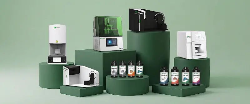
An open digital workflow provides maximum flexibility. High-accuracy intraoral scanners such as the Aident AI-30 deliver precise, true-color data suitable for soft-tissue monitoring. Compatible dental 3D printers (Ai-C60 or Ai220) and model resins then allow efficient production of educational and planning models when needed. Because all files use standard open formats, clinics retain full control of their data and can integrate with existing practice management or specialist systems.
Conclusion
Digital intraoral scanning combined with selective 3D printing offers a modern, efficient approach to periodontal monitoring and patient communication. Accurate, repeatable three-dimensional records improve clinical decision-making, while printed models enhance patient understanding and interdisciplinary collaboration.
Clinics that adopt this digital approach typically achieve better documentation, higher patient engagement, and more streamlined long-term care for periodontal patients.
Ready to modernize periodontal monitoring and model applications in your practice?
Explore Aident's open digital dentistry solutions-AI-30 Intraoral Scanner, dental 3D printers, and model resins-at Aident3D.com. Request a quote, book a live demonstration, or contact us via WhatsApp +86 19311417410 or email dongjiahao@aident.cc.